Optical Coherence Tomography
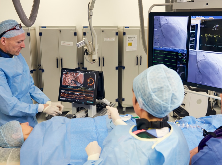
Optical coherence tomography (OCT) is an imaging technique used to visualize the inside of a coronary artery (blood vessel supplying the heart) using backscattered infrared light to generate images on a computer screen. OCT is usually done to guide a percutaneous coronary intervention (PCI) procedure. For example, an OCT may be used prior to implanting a stent to help the cardiologist measure the exact length and diameter of the narrowed segment of the blood vessel.
“My utmost priority is to provide the highest quality and most up-to-date cardiovascular care to all my patients. I aim to achieve this using a holistic approach in a personable and empathetic atmosphere, while taking into consideration each individual patient’s own wishes and hopes for their medical care.”
Using this information, the cardiologist can then choose the most appropriately sized stent. This enables a more individualized and tailored approach to coronary stenting and reduces future complications. In addition, OCT may be used after implanting a stent to ensure that the stent is properly implanted without any complications that may not be visible on the angiogram. During an OCT procedure a small catheter is inserted into the coronary artery over a wire. The coronary artery is flushed with contrast medium when taking the images in order to clear the blood from the artery. The imaging test itself takes only a few seconds to perform.